


Blog - October 19, 2026
What 2 Million Downloads Taught Us About The Usage of Foundation Models for Biology
From benchmarking to biomarker discovery, Bioptimus’s first model family, H‑Optimus has become part of research workflows we never expected to see, and it has taught us a lot about how general-purpose models create value.
In July 2024, just three months after launching the company, we released H‑Optimus‑0, our first foundation model for pathology, as an open-weight model on Hugging Face. Since then, the model has spread across universities, hospitals, pharmaceutical companies, biotech companies and AI pathology labs around the world. It has been used to compare models, automate routine pathology tasks, predict biomarkers, study patient outcomes and decide where expensive biological experiments should be run.
Throughout this piece, “H‑Optimus” refers to the three models we have made openly available to researchers: H‑Optimus‑0, released with open weights under an Apache 2.0 licence for anyone to use, including commercially; and H‑Optimus‑1 and H0‑mini, both available under a non-commercial research licence. We have written separately about why we chose to open the models; this is the story of what happened next.
From six downloads to 2 million
H‑Optimus‑0 had six downloads at the end of July 2024.
By the end of that year, it had passed 44,000. By March 2025, the H‑Optimus family had reached almost 175,000 downloads. Six months later, it had passed 800,000. At the end of 2025, it reached one million.
Today, H‑Optimus has been downloaded nearly 2 million times.
Figure 1: Cumulative H‑Optimus downloads. All H‑Optimus models combined, July 2024 to October 2026.
A Hugging Face download is not the same as a unique user. Models can be downloaded repeatedly when someone starts a new environment or runs a new workflow. So we see it as a measure of distribution and usage.
But downloads are only one part of the picture. We also have several thousand registered users, declared applications, publications and direct conversations with researchers. Taken together, they show something much more interesting than a successful launch spike: the models are being used repeatedly, across a large and growing number of organisations.
Adoption did not happen one institution at a time
One of the things we found was how adoption spreads.
Figure 2: Where H‑Optimus is used. Institutions in 64 countries around the world are confirmed to be using our models. (Gradient is for decorative purposes only)
We imagined that a university or pharmaceutical company might make a coordinated decision to use H‑Optimus. The reality was much more organic. One researcher tries it. Someone in a neighbouring team hears about the results. Another department starts benchmarking it. Eventually, we discover that multiple groups in the same institution have independently built it into their work.
At one university in Asia, 18 researchers use H‑Optimus; at another, it’s 14. At a US university, users span both its engineering and medical schools. The pattern is global: researchers are using H‑Optimus at institutions like MIT, Harvard, Stanford and Penn in the US; Oxford and Cambridge in the UK; Karolinska, ETH Zurich and Helmholtz Munich in continental Europe; HKUST, Tsinghua, Fudan and Seoul National University in Asia; universities in Brasília and Rio Grande do Norte in Latin America; and Mohammed V University and Babcock University in Africa.
Figure 3: H‑Optimus is used by well over 1,000 institutions worldwide. Some of the most renowned institutions using our models.
The same pattern appears inside pharmaceutical companies. H‑Optimus does not belong to one central “AI team.” Computational pathologists may use it as a feature extractor. Translational researchers may use it to investigate biomarkers. Clinical teams may explore patient stratification and outcome prediction. Infrastructure teams work out how to run it securely across large slide collections.
In some cases, institutions do not realise how widely the model is already being used internally until we show them. In fact, H‑Optimus has been accessed by more than 350 industry and healthcare organisations, including pharmaceutical and biotech companies, diagnostics and digital-pathology businesses, healthcare providers, biological-data companies, AI labs and hyperscalers. Adoption is often bottom-up, with multiple independent users appearing within the same organisation and double-digit user communities emerging inside some large pharmaceutical companies.
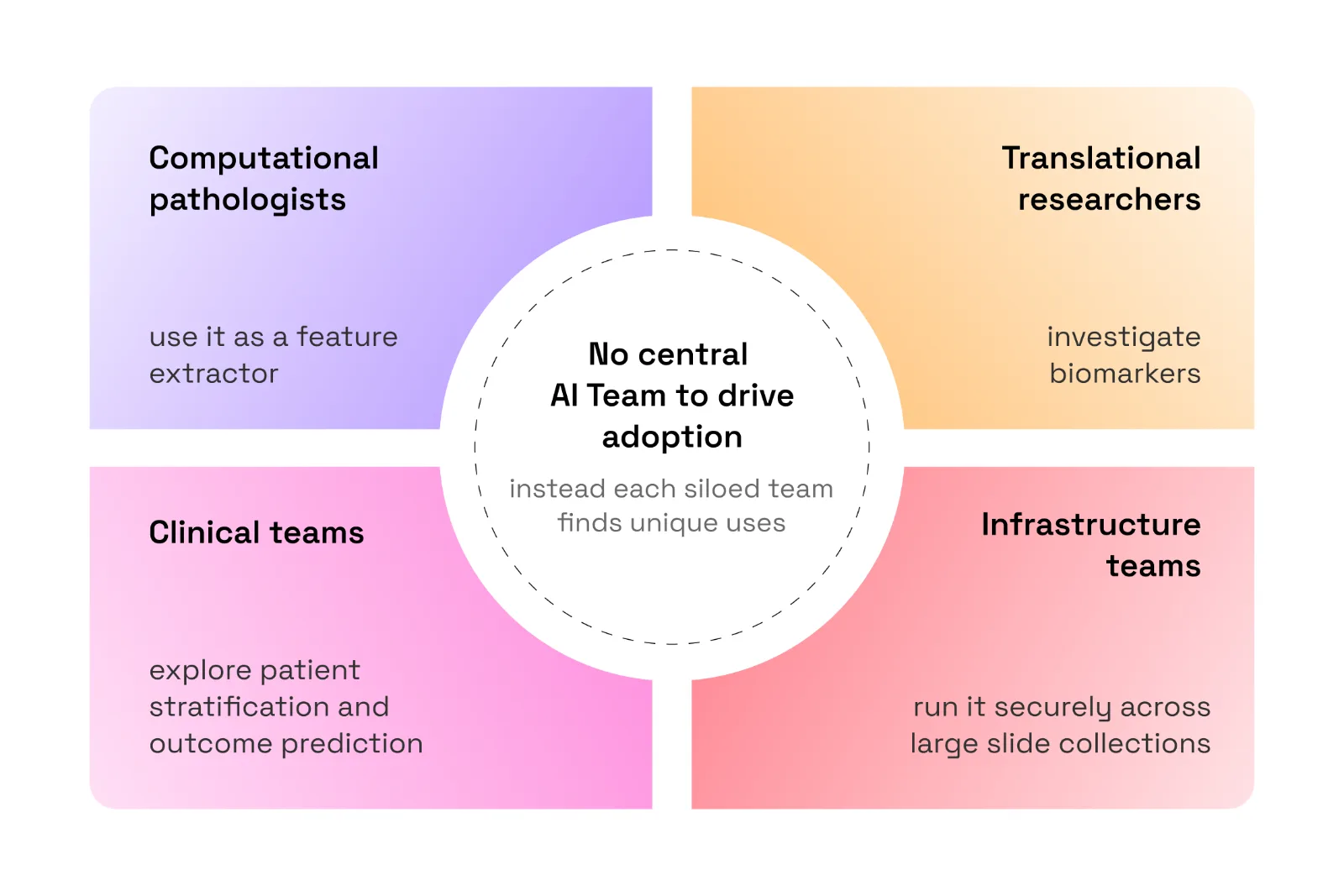
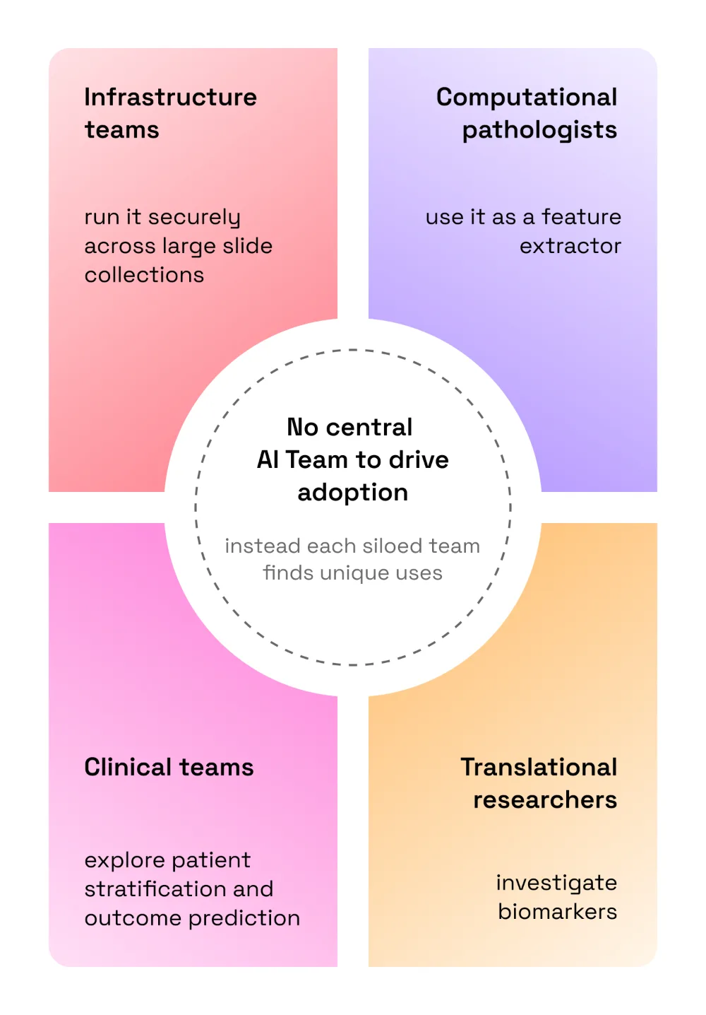
Figure 4: Inside one pharmaceutical company. We found that many companies do not have a central AI team yet; each siloed team finds its own uses for H‑Optimus.
That is both very cool and it tells us something about innovation and early adoption within companies. Adoption of open models is usually bottom-up. Institution-wide adoption comes later.
What are people using it for?
Of declared H‑Optimus applications, almost 60% began with model benchmarking.
Figure 5: H‑Optimus applications: Share of declared applications by type.
Benchmarking is the starting point. Researchers want to know whether a model works on their tissue, their scanner, their staining process and their patient cohort and not only on the data selected by its creators.
But rankings and comparisons are only the beginning.
Across the community, we now see H‑Optimus used for tissue and cell segmentation, slide quality control, region-of-interest selection, whole-slide aggregation and retrieval of morphologically similar tissue. Researchers are predicting mutations, biomarkers, gene signatures, tumour microenvironment features, recurrence risk, survival and treatment response.
Some teams use the embeddings directly. Others fine-tune the model or train task-specific heads on top of them. Others use H‑Optimus to help build an entirely new model, combining the general representations it has already learned with more data from a particular disease, institution or application.
Commercial value will vary widely by use case, but successful applications could each create millions of dollars in value and, collectively, point to a multibillion-dollar opportunity across diagnostics, research and drug development.
The breadth of these applications validates the value of general-purpose models: the same pathology representations can be reused across diseases and institutions, providing a foundation on which many different solutions can be built.
The range of indications
Applications span a wide range of diseases, with the strongest concentration in oncology. More than 95% of access requests that name a disease relate to oncology, but within cancer the work spans a broad spectrum of tumour types. Unspecified or pan-cancer applications form the largest group, followed by gastrointestinal and breast cancers, with substantial activity across thoracic, genitourinary, gynaecological and skin cancers as well as CNS tumours, haematological malignancies and sarcomas.
Figure 6: The range of indications. Based on explicit indication mentions in H‑Optimus model-access requests. Applications can mention more than one indication; non-human and cross-species work is identified separately from the request descriptions. Percentages may not total exactly 100% due to rounding.
The breadth does not stop at cancer, or even at human biology. Researchers are also applying H‑Optimus to inflammatory and immune-mediated disease, fibrosis, kidney disease, infectious disease and cardiovascular conditions. Others are testing it in mouse and rat models, comparative pathology and veterinary research. The table shows the distribution across indications; together, these applications suggest that a general representation of tissue morphology can travel across diseases, organs and species.
The oncology value propositions
Across oncology, the value propositions converge around four connected needs:
- Infer molecular biomarkers. Researchers use routine H&E to investigate mutation status, gene-expression signatures, MSI, HRD and immune markers.
- Predict outcomes and treatment response. Applications include prognosis, recurrence, survival and response to immunotherapy, platinum therapy and neoadjuvant regimens.
- Understand spatial biology. Teams study cell composition, tissue architecture and the tumour microenvironment.
- Accelerate core pathology workflows. H‑Optimus supports cancer detection, grading, subtyping and tumour segmentation.
Most projects begin as research or benchmarking, but they point towards a clear path from a general image encoder to disease-specific biomarker models, patient-stratification tools and potentially more efficient diagnostic and drug-development workflows.
Beyond oncology
The non-oncology work is smaller in volume but scientifically diverse:
- Inflammation and immunology. Researchers are studying inflammatory bowel disease, tissue inflammation and therapeutic response.
- Fibrotic disease. Applications include liver and lung fibrosis and the prediction of portal-hypertension-related parameters.
- Kidney disease. Teams are exploring kidney-biopsy analysis, chronic-kidney-disease prognosis and renal pathology classification.
- Infectious disease. Researchers are applying the models to tuberculosis slides.
- Cardiovascular disease. Early work includes atrial-fibrillation biopsy benchmarking.
Together, these projects suggest that representations learned from large-scale histology may be useful wherever tissue morphology contains signals about disease state, progression or response.
Cross-species and animal work
A small but distinctive group of requests is testing whether pathology foundation models transfer beyond human tissue:
- Mouse histology. Teams are evaluating H‑Optimus on mouse tissue and using whole-slide images to explore the prediction of biological age.
- Rat models. Researchers are extracting features from rat cerebellar sections for a neurodevelopment study.
- Comparative pathology. One project compares hepatocellular-carcinoma morphology across species.
- Veterinary applications. Researchers are studying equine uterine tissue and spontaneous diseases in animals.
These examples span preclinical models, comparative pathology and genuine veterinary applications; an early signal that a sufficiently general representation of tissue may travel across species as well as across diseases.
Figure 7: Beyond oncology and beyond humans. Areas outside cancer and outside human tissue where researchers apply H‑Optimus.
Three examples of usage
At MIT, researchers compared pathology foundation models to predict progression-free survival across more than 2,600 patients with breast, lung and colon cancer. H‑Optimus‑1 was the top-performing model, with time-dependent AUCs approaching 0.8. The most powerful thing is not only the result: choosing the right foundation model allowed the team to stop spending time on the backbone and move forward with the actual clinical research. Read the MIT case study.
At Oxford, researchers used H‑Optimus to predict spatial gene-expression signatures from routine H&E slides in inflammatory bowel disease. The goal was very practical: identify the most informative slides and regions before spending time and money on expensive spatial transcriptomics assays. Read the Oxford case study.
At a European medical centre, researchers used H‑Optimus‑1 as the foundation for a new diagnostic test in solid-tumour oncology. In external cohorts, the test achieved an AUC of 0.95 and showed the potential to reduce molecular testing by more than 75%. It is now being prepared for commercial deployment as a diagnostic. More to come on this very soon!
These examples show the breadth of high impact patient use-cases but also the potential for commercial opportunities to arise out of pioneering research based on open models.
What we have learned
1. Benchmarking matters, but it only measures what we already know to ask
Benchmarking was the most common declared use case, and for good reason. Independent comparison creates trust. H‑Optimus‑1 is currently ranked first and H‑Optimus‑0 third on PathBench, one of the most comprehensive evaluations in the field, covering 229 clinically relevant tasks.
But even 229 tasks capture only part of what these models can do.
Most established benchmarks were designed for familiar pathology problems: classification, detection, molecular prediction and survival. They are less able to evaluate the functionality emerging from newer models – connecting morphology to omics, combining different biological modalities, selecting the next experiment, adapting efficiently to a small proprietary dataset or becoming part of an agentic research workflow.
This is going to become a bigger issue. If a new model does not fit neatly into an existing benchmark, that does not necessarily mean it is less useful. It may mean the benchmark was designed for the previous generation of models.
We still need rigorous comparison. But we also need new ways to measure whether a model helps scientists ask better questions, generate new biological insight or reach an answer faster.
2. A general model creates value in lots of different ways
The range of applications has reinforced our belief in general-purpose foundation models.
The same model can provide embeddings for a segmentation workflow, support biomarker discovery, help predict patient outcomes or become the backbone of a more specialised model. With fine-tuning and additional data, it can be adapted to a specific disease or institution. Through inference, it can extract useful representations from slide collections that were previously very difficult to analyse at scale.
We see this reuse across gastrointestinal, breast, thoracic, genitourinary, gynaecological and skin cancers, as well as CNS tumours, haematological malignancies and sarcomas. Applications also extend beyond oncology to inflammatory and immune-mediated diseases, fibrosis, kidney disease, infectious disease and cardiovascular conditions and even outside of human biology.
That creates the potential for groundbreaking results. But we have also learned that the most valuable applications are not always the most glamorous.
Predicting a new biomarker is exciting. But helping a researcher choose the right 20 regions for an expensive experiment can save months. Automatically excluding unusable slides can unblock an entire data pipeline. A strong feature extractor can prevent every lab from having to rebuild the same foundational capability from scratch.
Often, the “boring” workflow problem is the one that determines whether ambitious science can happen at all.
3. Open usage showed us the unmet needs
We learnt where researchers repeatedly struggle: limited labelled data, expensive molecular assays, scarce linked clinical data, scanner and staining variation, compute constraints, and models that are technically available but still difficult to integrate.
We also saw the gaps in our own models. Performance is not equally strong across every tissue or application. Rare biological signals remain difficult. Researchers often need better tooling, documentation and deployment options as much as they need a better checkpoint.
Releasing the models did more than distribute our work. It gave us a much clearer map of what the field still needs.
4. Adoption spreads before institutions know it
Perhaps the clearest lesson is that a useful model disappears into the workflow.
It becomes a line in a pipeline, an encoder inside another model, a recurring step in an experiment or a shared tool passed between research teams. By the time an institution starts thinking formally about deployment, governance or infrastructure, the model may already be used across several departments.
That is how H‑Optimus has spread: not through one top-down decision, but through thousands of individual decisions by researchers who found it useful.
The 2 million downloads are one measure of that journey. The more meaningful measure is how much work now has H‑Optimus somewhere inside it, and what researchers have been able to build from there.
Where it all begins
None of this would exist without the patients. Every H‑Optimus model is built from tissue samples that people gave in the course of their own care, often at some of the hardest moments of their lives. Millions of these anonymised slides, carefully prepared and digitised by pathologists and hospital teams, are what we learn from: the variety in the patterns of human disease is what turns a neural network into a general representation of tissue. That representation now helps researchers in more than a thousand institutions find biomarkers, choose better experiments and design new diagnostics. We want to thank every patient whose data made this possible, along with the clinicians, pathologists and partner institutions who look after that data with care and consent. What started with them now returns to patients everywhere, as faster research, better diagnostics and medicines that reach the people who need them.
Ready To Get Started?
Off the shelf access. Evaluation support. Strategic collaboration.
Amazon
SageMaker AI
SageMaker AI
H-Optimus can be accessed through Amazon SageMaker, AWS’s fully managed ML platform, allowing teams to evaluate, fine-tune, and deploy the model within their existing cloud infrastructure.
Academic
Licenses
Licenses
Universities and research institutions can request non-commercial research licenses for evaluation and experimental use.
Industry
Licenses
Licenses
H-Optimus is available for collaboration with industry and clinical partners seeking to accelerate biomarker development, patient stratification, and translational research initiatives.
Not sure where to begin?
Read our full documentation on getting started: